
TR-FRET Assay Principle
What is TR-FRET?
TR-FRET (Time-Resolved Fluorescence Resonance Energy Transfer) combines time-resolved fluorescence (TRF) detection with fluorescence resonance energy transfer (FRET) to provide sensitive and homogeneous detection of molecular interactions and biological analytes. TR-FRET assays can be used for the detection and quantification of proteins, cytokines, hormones, antibodies, and other biomolecules in samples such as serum, plasma, and cell culture supernatants.
The TR-FRET assay principle is based on energy transfer from a fluorescent donor to a fluorescent acceptor when the two fluorophores are brought into close proximity through a specific molecular interaction. Following excitation of the donor, energy transfert to the acceptor results in acceptor fluorescence. The fluorescence is measured after a defined time delay, taking advantage of the long-lived emission of lanthanide-base donors. This time-resolved detection helps reduce short-lived background fluorescence, supporting sensitive and reliable quantitative analysis.
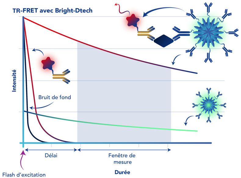
In the Bright-Dtech™ TR-FRET format, detection antibody is coupled to a Bright-Dtech™ fluorescent lanthanide nanoparticle acting as the donor, while a second detection antibody is coupled with fluorescent molecules acting as the acceptor. When both detection antibodies bind to the same target, the donor and the acceptor are brought into close proximity, enabling FRET. The resulting TRF signal can be measured using a compatible TR-FRET microplate reader.

TR-FRET can be applied to a wide range of research applications, including biomarker detection, protein–protein interaction studies, drug discovery, molecular screening, and biochemical research.
Required material : Black microplates, plate shaker, and compatible TR-FRET microplate reader.
TR-FRET vs conventional FRET: What is the difference?
Both FRET and TR-FRET are based on energy transfer between a donor and an acceptor when they are in close proximity. The main difference is the fluorescence measurement approach.
In conventional FRET, fluorescence is typically measured immediately following excitation. As a result, fluorescence originating from the sample, plate, or other assay components may contribute to the measured signal.
In TR-FRET, fluorescence is measured after a defined delay following excitation. This takes advantages of the longer emission lifetime of lanthanide-based donors, allowing short-lived background fluorescence to decay before measurement. As a result, TR-FRET can provide improved signal-to-background performance and sensitive detection in complex samples.
TR-FRET Protocol
The Bright-Dtech™ TR-FRET workflow uses a simple homogeneous mix-and-read format. Samples or standards are combined with the donor- and acceptor-labelled detection reagents and incubated to allow formation of the detection complex. Following incubation, the fluorescence signal is measured using a compatible TR-FRET microplate reader.
No washing or separation steps are required, providing a simple workflow with reduced hands-on time.

Benefits of TR-FRET with Bright-DtechTM
Bright-Dtech™ fluorescent lanthanide nanoparticles provide high signal intensity, long-lived fluorescence, and excellent photostability for TR-FRET applications. Combined with time-resolved detection, these properties support:
- High signal intensity for sensitive detection.
- Low background fluorescence through time-resolved measurement.
- Excellent signal-to-background performance.
- Long-lived fluorescence enabling time-resolved detection.
- High photostability for reliable fluorescence measurements.
- Homogeneous, wash-free assays with simple mix-and-read workflows.
- Reduced hands-on time compared with separation-based assay formats.
Example: TR-FRET Assay for Human CRP Detection
C-reactive protein (CRP) is an acute-phase protein widely used as a biomarker of inflammation. Sensitive and quantitative CRP detection can support research into inflammatory responses and disease-related processes.
The Now-Dtech™ Human CRP Assay Kit provides a homogeneous, wash-free approach for the quantitative detection of human CRP.
For CRP detection using the Bright-Dtech™ TR-FRET format, samples or standards are added to the microplate wells together with donor- and acceptor-labelled detection reagents. The detection antibodies bind to CRP, bringing the donor and acceptor into close proximity and enabling FRET. Following incubation, the TRF signal is measured using a compatible TR-FRET microplate reader.
Within the assay range, the measured signal increases with the concentration of CRP present in the sample, allowing quantitative determination using a standard curve.
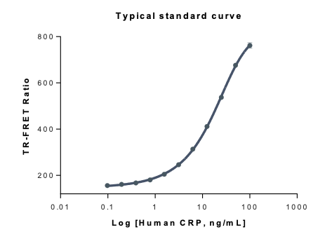
Figure 1. Human CRP detection using TR-FRET assay principle
Bright-Dtech™ technology supports sensitive human CRP detection through the combination of high signal intensity, long-lived lanthanide fluorescence, and time-resolved detection.
Assay range: 600-100,000 pg/mL
LOD: 110 pg/mL